Specialised edition developed with advice and guidance from the
Thomas Pocklington Trust
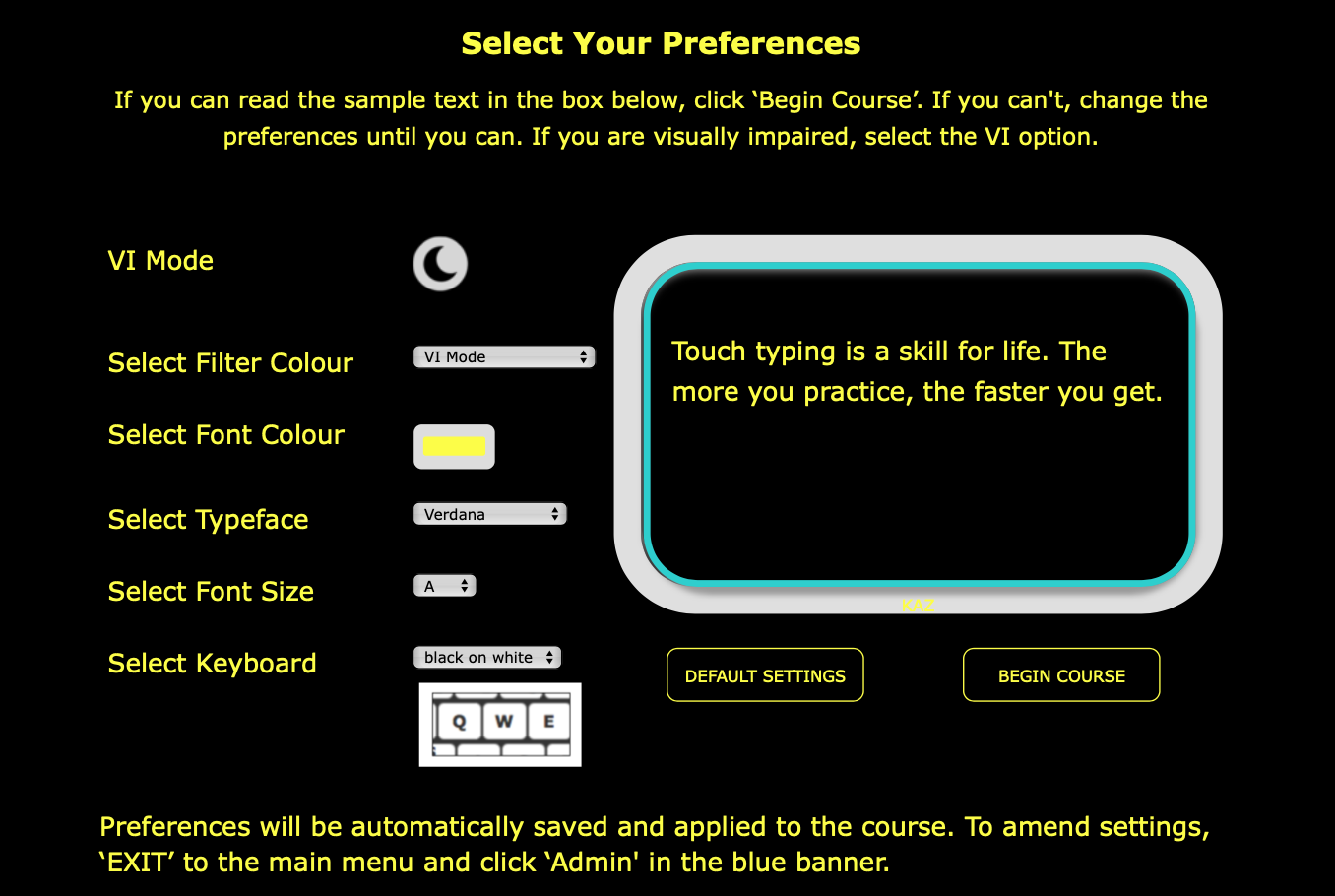
Compatible with:
JAWS and other screen readers
Dolphin SuperNova and other magnification software/hardware
Google and other captioning software
Learning to touch type is considered one of the most beneficial skills for visually impaired and blind individuals. This is because it allows them to transfer their thoughts easily and automatically onto a screen. It provides them with an invaluable tool and asset for independent working and communicating.
Learning to touch type at any age can dramatically boost confidence, self-belief and independence. However, teaching learners with visual impairment at an early age can drastically transform their experience whilst at school and in FE/HE. It puts them on a more even standing with their sighted peers and opens doors to new career opportunities.
Achieving muscle memory and automaticity when touch typing increases efficiency and productivity. However, most importantly, it frees the conscious mind to concentrate on planning, composing, processing and editing, greatly improving the quality of the work produced.
Neuroradiology typically categorizes pathological findings into three major anatomical regions. 1. The Brain
Uses ionizing radiation to create cross-sectional images based on tissue attenuation, measured in Hounsfield units (HU).
Neuroradiology: The Essentials with MR and CT Neuroradiology is a specialized field of medical imaging focused on diagnosing disorders of the brain, spine, and head and neck. Central to this discipline is the strategic use of and Magnetic Resonance Imaging (MRI) , which provide the high-resolution visualization necessary for modern neurological care. The Core Modalities: CT vs. MRI
CT angiography (CTA) is frequently used to assess vascular issues like aneurysms and acute stroke. Magnetic Resonance Imaging (MRI):
Ideal for initial evaluation in emergency settings due to its speed and widespread availability. It is excellent for detecting acute hemorrhage, bony fractures, and calcifications.
Techniques like diffusion-weighted imaging (DWI) are critical for early stroke detection, while spectroscopy and perfusion imaging provide metabolic and functional insights. Key Clinical Areas of Focus
Employs powerful magnetic fields and radiofrequency pulses to capture signals from water and organic molecules.
Offers superior soft-tissue contrast resolution, making it the preferred choice for detailed analysis of tumors, inflammation, and chronic conditions. It is highly sensitive for white matter diseases like multiple sclerosis.
Neuroradiology: The Essentials With Mr And Ct Now
Neuroradiology typically categorizes pathological findings into three major anatomical regions. 1. The Brain
Uses ionizing radiation to create cross-sectional images based on tissue attenuation, measured in Hounsfield units (HU).
Neuroradiology: The Essentials with MR and CT Neuroradiology is a specialized field of medical imaging focused on diagnosing disorders of the brain, spine, and head and neck. Central to this discipline is the strategic use of and Magnetic Resonance Imaging (MRI) , which provide the high-resolution visualization necessary for modern neurological care. The Core Modalities: CT vs. MRI Neuroradiology: The Essentials with MR and CT
CT angiography (CTA) is frequently used to assess vascular issues like aneurysms and acute stroke. Magnetic Resonance Imaging (MRI):
Ideal for initial evaluation in emergency settings due to its speed and widespread availability. It is excellent for detecting acute hemorrhage, bony fractures, and calcifications. Neuroradiology: The Essentials with MR and CT Neuroradiology
Techniques like diffusion-weighted imaging (DWI) are critical for early stroke detection, while spectroscopy and perfusion imaging provide metabolic and functional insights. Key Clinical Areas of Focus
Employs powerful magnetic fields and radiofrequency pulses to capture signals from water and organic molecules. MRI CT angiography (CTA) is frequently used to
Offers superior soft-tissue contrast resolution, making it the preferred choice for detailed analysis of tumors, inflammation, and chronic conditions. It is highly sensitive for white matter diseases like multiple sclerosis.